About Eye Anatomy
We think that everyone has the right to see the world accurately. That’s why we’ve chosen to focus on delivering the greatest eye care possible.
We All Deserve To See The World with Clarity.
The human eyes is one of the most delicate and treasured parts of the body. Blindness and vision impairment are the primary concerns for most people since eyes are responsible for viewing the beauty of nature around us. Our eyes allow us to see and differentiate between different colors, forms, and sizes. However, the eyes can also be affected by various diseases and disorders which may lead to vision impairment or even blindness.
The eye is the medium between our surroundings and our brains. It converts light rays into messages to the brain, which are then encoded into visual images by the brain. The eye’s 5 basic components, each measuring one inch in diameter and placed in a protective cone-shaped chamber within the skull, play an important part in vision development. The eyeball is the white, round, firm structure that houses the other four eye parts. The clear, dome-shaped surface at the front of the eye is called the cornea. It protects the iris (the colored part of the eye) and pupil (the black center of the eye) from dirt and debris.
Book Free Consultation
Book Appointment or Video Consultation online with top eye doctors
Eye Anatomy
The eye’s basic and fundamental anatomy comprises of:
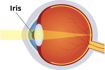
Iris
The iris is the transparent front part of the eye, which contains different colors for different people. The pupil is in the middle of the iris and acts as a control mechanism for controlling the size of the pupil during relaxation or expansion process. The color of the iris is due to the pigmentation of the stroma, which is a thin layer of tissue in the eye. The pigmentation can be brown, blue, green, hazel or any other color.
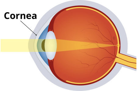
Cornea
The cornea, or outer layer of the eye, covers the anterior chamber. It is transparent and enables focusing power when light enters the eye. Tear fluid delivers nutrients and oxygen to the five-layer structure. The outside layer is made up of epithelium, which contains high regenerative cells that aid in rapid recovery from shallow wounds. The middle layer consists of stroma, which is the thickest and most central layer of the cornea. This layer provides strength to the cornea. The innermost layer is endothelium, a single cell layer that helps regulate fluid balance within the stroma. The clearness of the cornea is essential for good vision. It can become cloudy due to injury, disease, or inherited conditions.
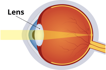
Lens
The lens is located directly behind the iris and is a clear and transparent structure. The lens’s primary function is to alter its shape from thick to thinner or vice versa, focusing for near and long vision. When the eye is at rest, the lens is flat and when we look at something near, the lens becomes more convex (round). The ciliary muscles are tiny muscles attached to the outside of the eye that help the eye focus by changing the shape of the lens.
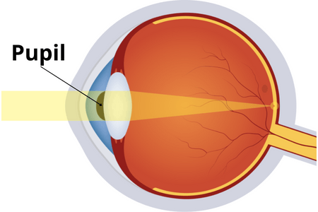
Pupil
The pupil is the black center of the iris that absorbs most of the light as well as controlling how much light enters the eye. It is black in color because it absorbs all of the light that comes into the eye and then turns red in photos due to reflection from the retina. It constricts light passing through by restricting its brightness, then the iris adjusts the amount of light by expanding or contracting the pupil.
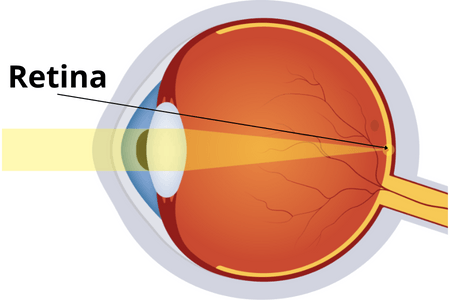
Retina
The light-sensitive tissue within the eye ball, known as the retina, is made up of delicate nerve fibers and two types of light receptors that are embedded inside the wall. This is color sensitive and contains rods that absorb black and white light. This serves as a film in a camera before being transmitted to the brain.